Which Leads Are Best for a Rhythm Strip
32 Atrial flutter basic Atrial Tachycardia with block if more advanced. When viewing the EKG strip V4-V6 on the strip will be referred to as V-13-15.
This is great for identifying baseline cardiac rhythm as well as any arrhythmias or ectopy that may occur like a premature beat.

. The first step in analyzing an EKG or ECG strip is to calculate the heart rate. A sinus morphology is an upright P wave in lead II and biphasic up and down P wave in lead V1. The goal of reading an EKG rhythm strip is to determine the rate and rhythm of the patient.
Any Clues As To Svt Diagnosis. Printing a longer strip called a rhythm strip gives the nurse enough QRSs to assess the rate and the rhythm. In these cases is possible to determine sinus rhythm because P waves fulfill the first three criteria described above replacing R-R interval with P-P interval.
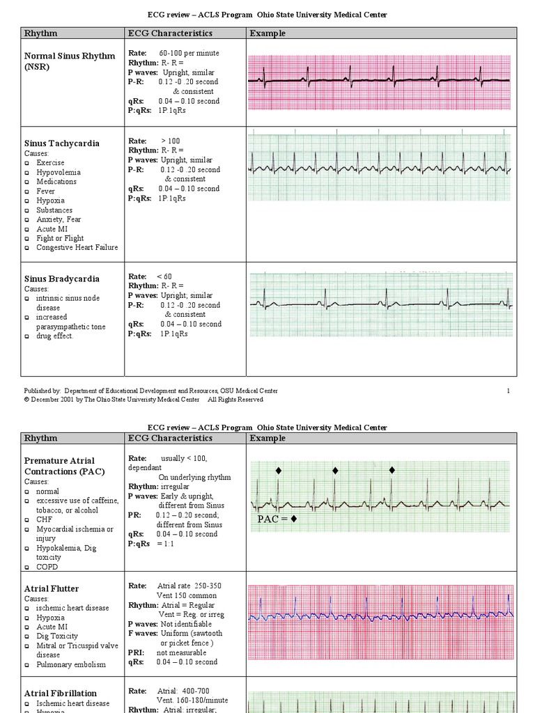
The best lead view on the monitor to examine is lead II. Cardinal features of sinus rhythm The P wave is upright in leads I and II Each P wave is usually followed by a QRS complex. The first ECG strip below shows a P wave with sinus.
Most rhythm strips are interpreted from Lead II as this gives a great view of the heart. Since an Apple Watch rhythm strip is only 1 lead and since it is done by the patients initiation and not. The interval between the P wave and each QRS complex is usually less than 02 seconds and should be consistent.
The usefulness of recording all 12-leads as a rhythm strip allows identification of paced complexes in all ECG leads. Once we understand what each part of the wave represents we can apply some simple steps to analyze the rhythm. This is the CPT code for interpretation of a 12-lead EKG if someone else usually the hospital owns the EKG machine.
Positive P wave in lead II and negative in lead aVR. What is v1 and v2 in ECG. It requires an order from a physician and a written interpretation.
The precordial or chest leads V1V2V3V4V5 and V6 observe the depolarization wave in the. Negative in leads aV R and V 1 to V 2. The EKG strip can be used to measure your hearts patterns for a full minute or even longer.
A complete three-dimensional electrical activity can be recorded using a 12-lead ECG which is a standard recommended assessment tool for diagnosing myocardial ischemia and infarction3 The rhythm. Home ECG Library ECG Basics ECG Basics Homepage The rhythm is best analyzed by looking at a rhythm strip. Heart rate between 60 and 100 bpm.
Lead II which usually gives a good view of the P wave is most commonly used to record the rhythm strip. In this manufacturers magnet mode 3 AV outputs are delivered at 100 bpm and short AV interval followed by outputs delivered at 85 bpm at the programmed AV interval designed to evaluate native AV conduction. Most schools do not spend an enormous amount of time.
Explain why the different waves of the ECG are seen as an upward deflection in some leads but a downward deflection in others. The PR interval should be calculated and consistency assessed throughout the rhythm strip. And biphasic in lead V 3.
Introduction Lead II is frequently unrepresentative of QRS configurations especially for arrhythmias. To assess the cardiac rhythm accurately a prolonged recording from one lead is used to provide a rhythm strip. 34 Sinus arrhythmia basic Sinus rhythm with blocke PAC if more advanced.
31 Second Degree AV Block Type I Wenchebach PR longer longer dropped beat. A longer rhythm strip recorded perhaps recorded at a slower speed may be helpful. There are different ways to calculate ECG heart rate on a 6 second strip.
Which leads are best for a rhythm strip. 33 Atrial FibrillationFlutter Goes between no discernible Ps and flutter waves. Aside from a 12-lead ECG placement theres something known as a 15-lead placement which includes placing leads V4-V6 on the posterior side of the patient below their left scapula see below.
The lead we are most familiar with is Lead II which is one of the limb leads. Confirm or corroborate any findings in this lead by checking the other leads. P-P interval must be constant.
To assess the cardiac rhythm accurately a prolonged recording from one lead is used to provide a rhythm strip. This can be done if a heart rhythm is. Remember that the QRS complex represents intraventricular conduction time.
Here there is atrial bigeminy due to. Why is a Rhythm Strip necessary. Positive with most of the complex above the baseline in leads I II III aV L aV F and V 4 to V 6.
For new students learning about the EKG process and EKG rhythms the information can be overwhelming. CPT code 93010 Medicare reimbursement about 850. EKG rhythm strips are generally used when the normal EKG does not produce desired results.
Lead I Lead II and Lead III combine to form a triangle around the perimeter of the heart. On a 12 lead ECG this is usually a 10 second recording from Lead II. Lead II which usually gives a good view of the P wave is most commonly used to record the rhythm strip.
How does atrial activity relate to ventricular activity. - Identify Rhythm Cardiac Abnormalities - Provides good view of P Wave. The best lead for observing atrial activity is lead II.
One of the easiest ways to calculate heart rate on a 6 second strip is to count the amount of R waves on a 6 second strip and and multiply it by 10. This triangle is frequently referred to as Einthovens Triangle named in the early 1900s after a pioneer in electrocardiography.

12 Lead Ecg Interpretation Lesson And Practice Quiz 311 Ekg Interpretation Ecg Interpretation Emergency Nursing

08 Read Ecgs Like An Expert Basic Ekg Interpretation Ekg Interpretation Nurse Quotes Ekg Interpretation Cheat Sheets

Ecg Learning Center An Introduction To Clinical Electrocardiography P Wave Learning Centers Tutorial Sites

Ecg Interpretation Cheat Sheet Ecg Interpretation Ekg Interpretation Cheat Sheets Ekg Interpretation

Best 12 Lead Ekg Interpretation Cheat Sheet Video Ever Created Ekg Interpretation Cheat Sheets Ekg Interpretation Ekg

Stemi Vs Nonstemi Emergency Nursing Medical Surgical Nursing Cardiology Nursing

Screen Shot 2018 07 29 At 10 22 00 Png Medical School Studying Cardiac Nursing Nursing School Essential

Ekg Examples Free Download As Pdf File Pdf Text File Txt Or Read Online For Free Cardiac Nursing Ekg Interpretation Cheat Sheets Ekg Interpretation

Rosh Review Medical Mnemonics Heart Blocks Medicine Notes

Pin On Nursing A Vocation Not A Job

Pin By Vivian Nwankpa On Emt Cardiac Nursing Nclex Nursing School Survival

Youtube Ekg Interpretation Heart Blocks P Wave

Ekg Examples Free Download As Pdf File Pdf Text File Txt Or Read Online For Free Cardiac Nursing Ekg Interpretation Cheat Sheets Ekg Interpretation

View In Full Resolution Ekg Interpretation Ecg Rhythms Ekg Rhythms

Cardiac Assessment Back Sheet Cardiacworkout Cardiac Assessment Emergency Nursing Nurse

Ecg Interpretation 25 Lead Vi Ecg Interpretation Ekg Cardiology Study


Comments
Post a Comment